Advantage of Quantitative Computed Tomography - pQCT
pQCT - Advantages over DXA and QCT methods
pQCT vs. DXA
With pQCT it is possible to distinguish between cortical and trabecular bone. Due to its larger surface area, trabecular bone reacts faster to changes in bone metabolism than cortical bone. Bone resorption or the success of a therapy can be detected earlier and with higher significance with pQCT than with methods that cannot distinguish between cortical bone and cancellous bone.

Physically correct density
(volumetric BMD, material properties)
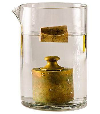
In contrast to the DXA method, the pQCT measures the actual density in g/cm³ and not the area-projected mass in g/cm².
When projecting the mass onto a surface, the information about the 3rd dimension of the bone is lost. It is thus no longer possible to distinguish between density and, for example, thickness. Therefore, small people are often wrongly diagnosed as osteoporotic because they have a lower bone mass. Especially in children and adolescents, it is therefore not possible to distinguish growth from an actual increase in bone density. Many children with growth retardation due to chronic disease have therefore been treated for osteoporosis. In contrast, pQCT can distinguish between size and density, since bone density as a material property is independent of size.

Bone geometry
With pQCT, not only the density but also the bone geometry can be measured. Bone strength depends on the spatial distribution of the bone material. From the cross-section geometry, the pQCT software can calculate the moment of inertia and the section modulus. These mechanical properties allow the prediction of the fracture load with high accuracy. Analysis of the cortical bone in the diaphysis can also distinguish osteomalacia from osteoporosis. Cortical density is normal (1100-1200 mg/cm³) in patients with osteoporosis, while patients with osteomalacia have decreased < 1000 mg/cm³) cortical density.

Highly reproducible measurements
Most DXA measurements are taken at the spine. With increasing age, the number and severity of degenerative changes in the spine increases, leading to falsely high values. As a result, an increase in degenerative changes can be misinterpreted as a therapeutic success. Therefore, it is recommended not to measure the spine if degenerative changes are visible in the X-ray.

Minimisation of measurement errors
Measurements using DXA or QCT are influenced by changes in the fat content of the bone marrow. While essentially only fatty marrow is found at peripheral measurement sites from the age of 20, the composition of the marrow in the spine and proximal femur can change considerably. The soft tissues surrounding the bone can also affect the result. For follow-up measurements, a change in fat content can alter the DXA results by up to 11%.

Muscle & bone diagnostics in one step
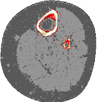
The pQCT measurement allows the comparison of muscle and bone parameters. The bone strength is adapted to the maximum muscle strength. With pQCT it is possible to determine both muscle cross-sectional area and muscle density. By comparing the muscle and bone cross-sectional area, it is possible to see whether the bone is adapted to the muscle. This allows to differentiate osteopenia due to muscle weakness from primary osteoporosis. In the first case, the low bone strength is the result of the physiological adaptation process of the bone to the low muscle strength. IIn this case, the bone is healthy and the muscle needs to be treated. In the second case, the bone is not adapted to the muscle and the bone must be treated.

Do you have questions about our products or would you like to purchase a product?
Let our experts advise you. Simply arrange a non-binding consultation.
Let Stratec pQCT convince you!
Numerous scientific studies prove the validity of Stratec pQCT. Benefit from over 30 years of experience in muscle and bone research and over 1000 scientific publications on pQCT worldwide.